New 3D camera for cleft surgery analysis
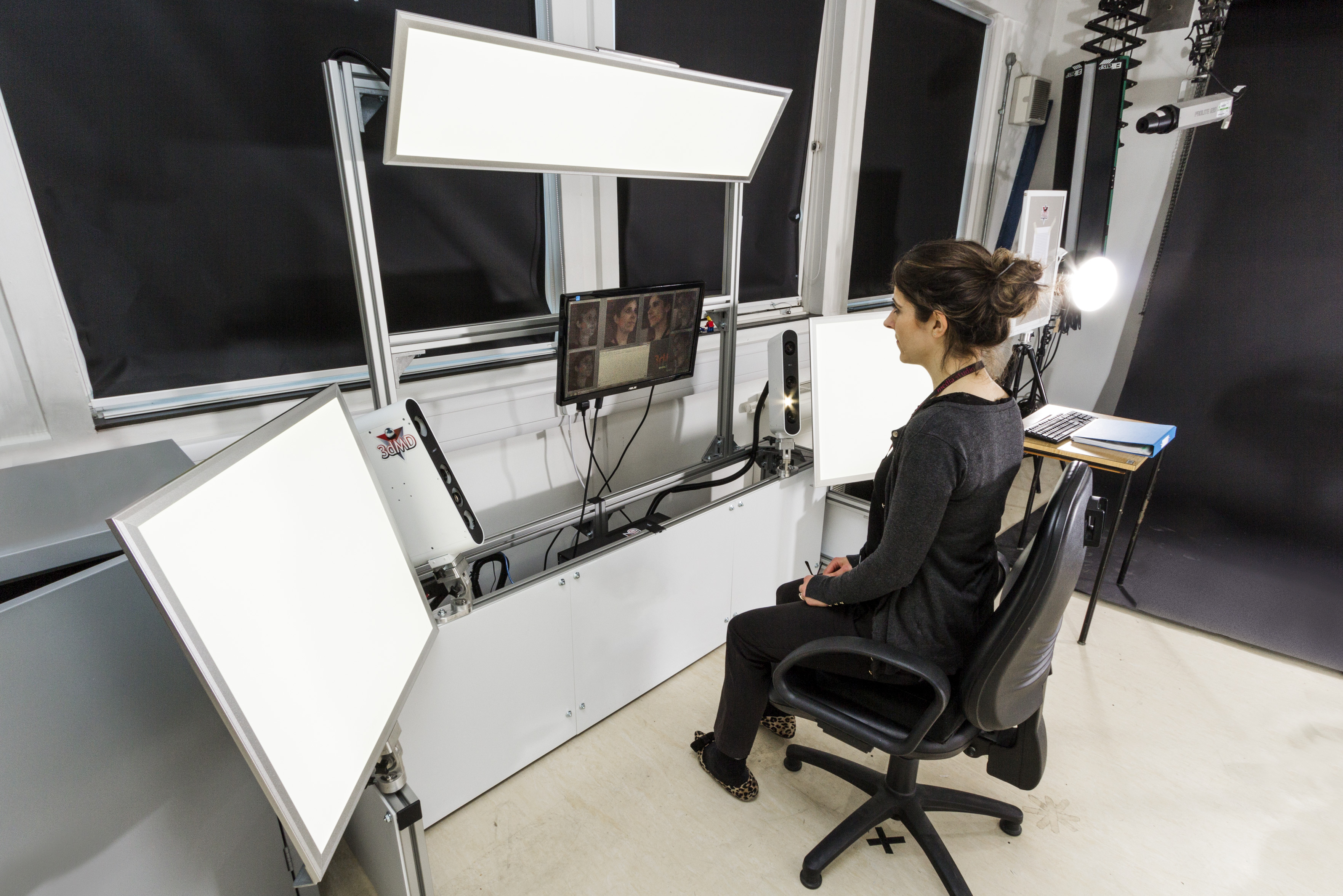
 Media Studio's clinical photography team has been recording 3D images of patients for over 20 years, comparing pre- and post-operative images of cleft and plastic surgery patients, but in that time the technology has moved on to a new level of precision. This week, we moved into a new era of 21st century imaging, commissioning a 3D camera that belongs to a completely new generation of equipment.

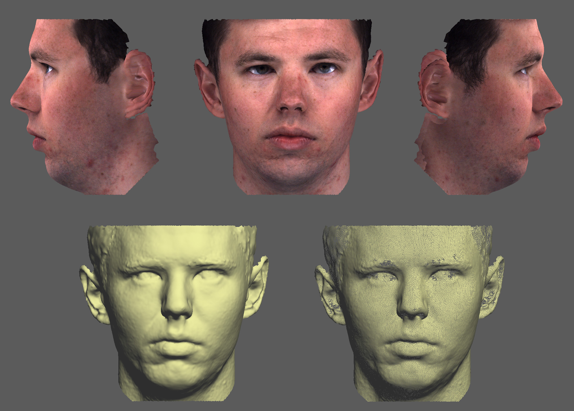
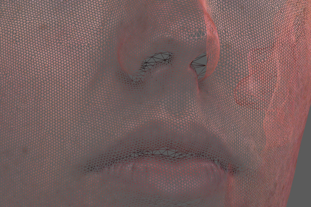
The new camera can capture seven frames per second, enabling us to record moving 3D images and allowing us greater choice and flexibility in the selection of still images. This is a very useful feature for recording small children – including new-born children with cleft lip and palate – making it easier to get the correct patient positioning for analysis. Where the old system used flash to light the patient, the new camera has broad, continuous-lighting panels, which results in a more evenly-lit image with minimal shadowing. This type of camera works by recording separately the precise geometry of the face and its surface texture to render the image in full colour. This new camera gives us approximately double the resolution in both images, as well as the ability to analyse both static and moving 3D images.

3D imaging is currently being used for a range of studies, including:
- Dynacleft study: looking at the way Dynacleft webbing changes the size and shape of the premaxilla in bilateral cleft lip patients, with the aim of reducing surgery from two stages to one stage.
- Autism facial morphology: looking at the connection between facial morphology and autism using 3d analysis and machine learning.
- Cleft Collective: part of a study which aims to find the causes of cleft by looking at genetics, family history, substance use and all other metrics that could contribute.
- Haemangioma: looking at haemangioma growth over time using 3D volume and surface area analysis.
